
Ultrasound
Real-Time Diagnostic Imaging
Ultrasound uses high-frequency sound to create detailed images of soft tissues, organs, and blood flow. It's safe, painless, and requires no radiation exposure.
Key Features
- Real-time imaging of organs and soft tissues
- Evaluation of blood flow using Doppler technology
- Noninvasive imaging without ionizing radiation
Commonly used to evaluate
- Blood flow and vascular blockages
- Narrowing of blood vessels
- Tumors and congenital abnormalities
- Organ structure and function
What to Expect
Preparing for Your Exam
Preparation for an ultrasound exam is typically minimal. For some exams, such as those involving the gallbladder or abdominal organs, you may be asked to avoid eating or drinking (except water) for six to eight hours prior to the appointment. Specific instructions will be provided at the time of scheduling if required.
During Your Exam
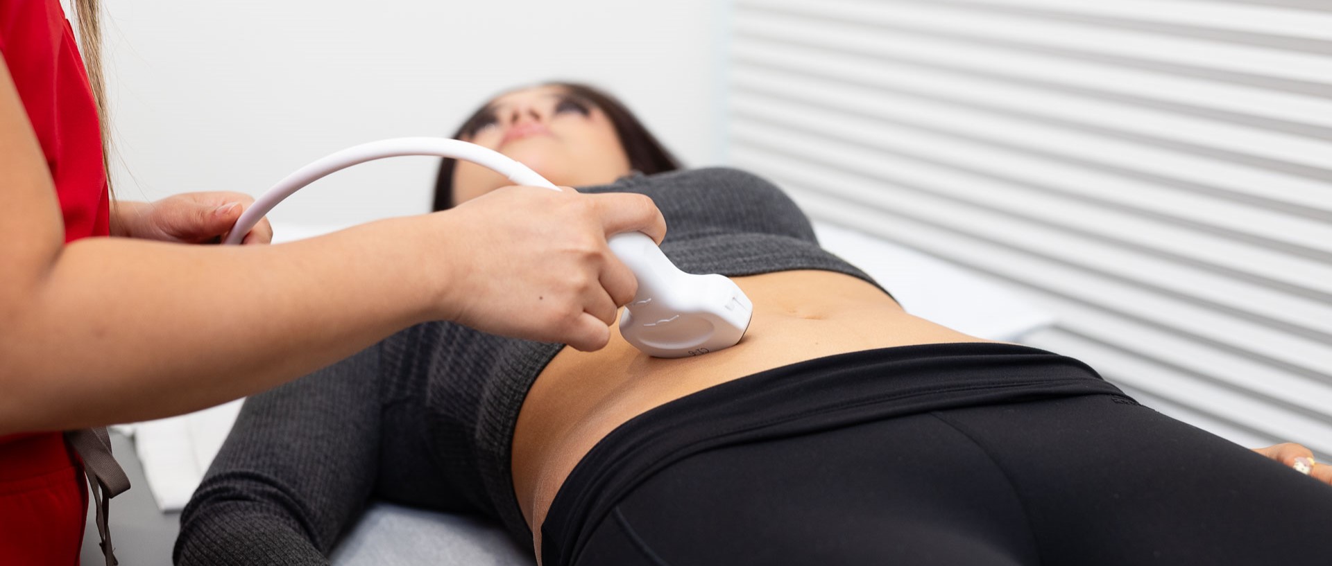
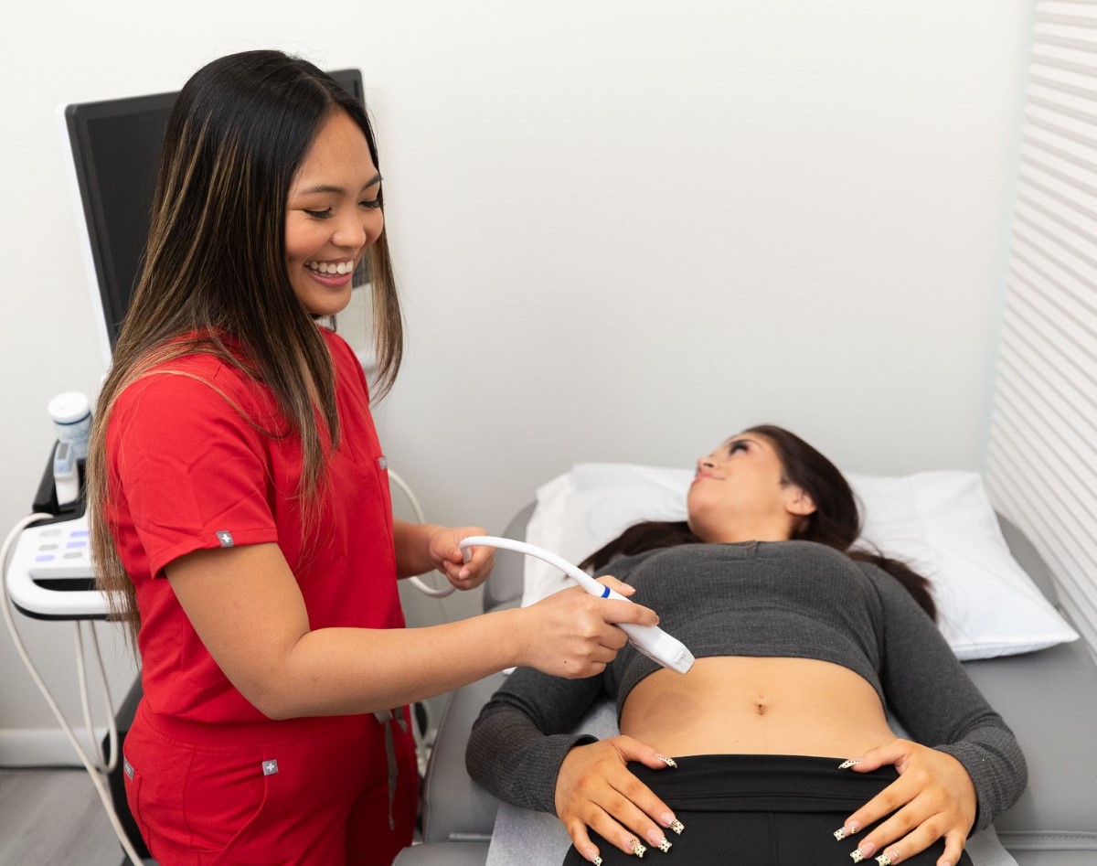
You may be asked to change into a gown before the exam. During the procedure, you’ll lie comfortably on an exam table while a technologist applies a small amount of gel to the area being examined. The gel helps transmit sound waves and allows smooth movement of the transducer across the skin.
Ultrasound exams are painless, though mild pressure may be felt as the transducer is guided over the area. You may be asked to adjust your position to improve imaging. Once the exam is complete, the gel is removed, and normal activities may be resumed immediately.
Schedule Your Imaging Appointment
Appointments are available for a range of imaging services. Contact the office to schedule your visit.
